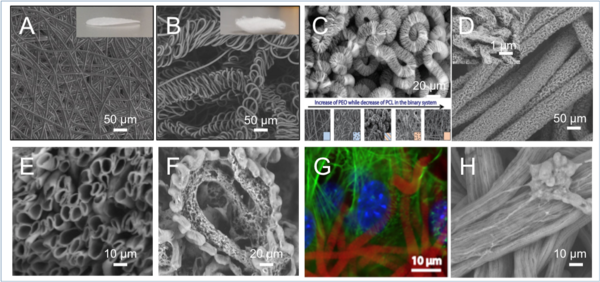
Conventional electrospinning normally generate 2D straight fibers (A) with limited cellular penetration. Here we explore new possibilities to upgrade electrospinning technology into 3D (B) and apply it 1) to fabricate 3D Nano-biointerfaces that recapitulate the 3D in vivo environment, 2) to guide 3D cell‐materials interactions and 3) to realise a wide range of biomedical engineering applications such as tissue engineering, biosensing and cancer biology.
We have discovered for the first time that a nanoscale immiscible polymer blend solution electrojet can assemble into ultraporous interweaving microfibers (C). This intriguingly novel morphology originated from a blend of polycaprolactone (PCL) and polyethylene oxide (PEO) in a DCM–DMF mixed solution when the ratio between each component reached a threshold and when the electrospinning parameters were delicately controlled. 3D microfibers with nanopores (D), core-shell (E), side-by-side (F) compartments have been developed for cell interactions (G: NIH3T3, red:fiber C, blue:nucleus, green:actin; H: PC12).